 Nitya Nandkishore The ability to bite, chew, swallow, wink, smile or frown comes from the muscles of the face and head. It is clear that what these muscles do is different from the muscles of the arm, leg or body, which help in picking up something, moving from one place to another or sitting straight. But all of these muscles develop from the same group of cells called the mesoderm. Then how do they learn what their roles in the adult are going to be?
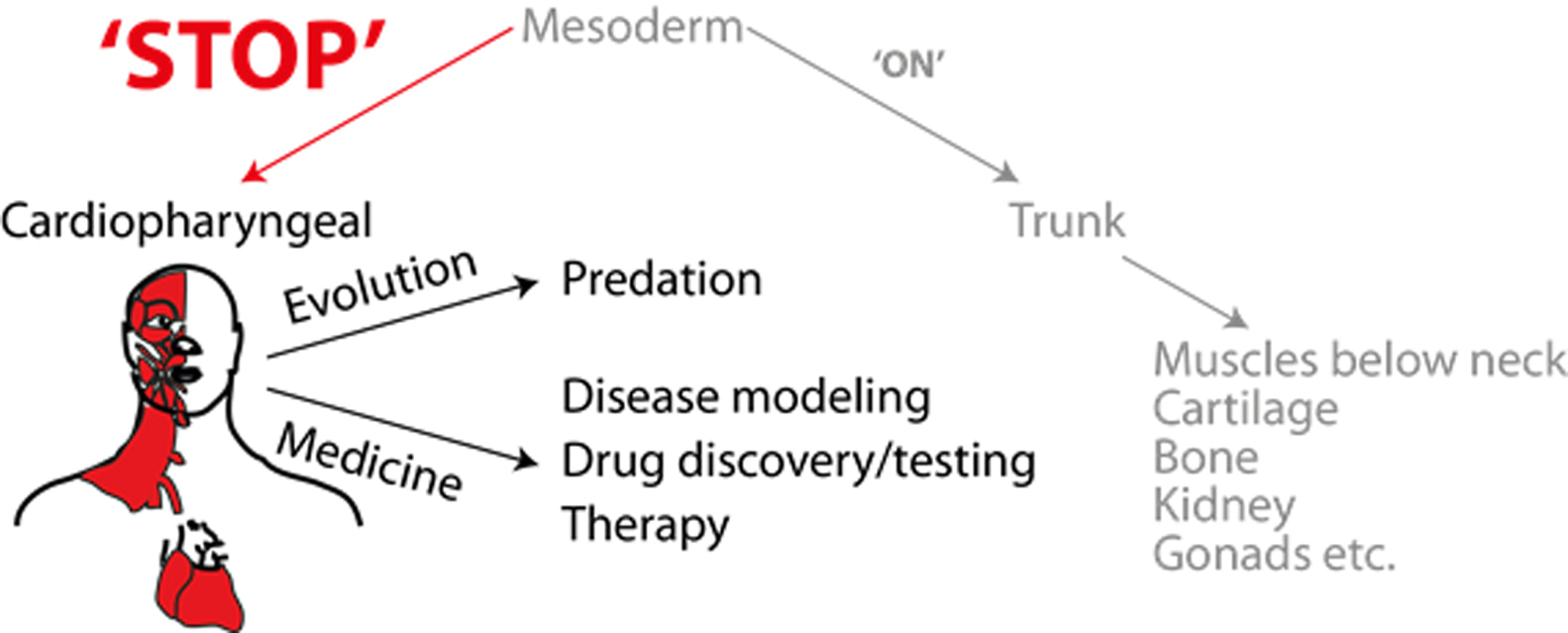
The single egg cell when fertilized by the sperm begins a series of divisions to form the important systems of the body. This period is called embryonic development. An important event in this period is gastrulation, the formation of the three germ layers – the ectoderm, the mesoderm and the endoderm. The ectoderm forms the nervous system and skin, while the endoderm forms the digestive system, lungs, liver and pancreas, as well as the internal lining of some systems. The mesoderm, or middle layer, is an extraordinarily versatile layer that forms the skeleton of the body, including bones, tendons, ligaments, as well as heart, kidneys and gonads (that produces the eggs or sperm). It is this germ layer that also forms the muscles of the body. So then how do the mesodermal cells make the choice to form muscles of the head or those below the neck? Previous studies have shown that these two types of muscles follow different routes to the end point of making muscle. All muscle ancestor cells start as mesoderm, become different at a point when they turn on separate sets of instructions, i.e., genetic programs, to specify them as head or trunk category and then again follow a common set of instructions to make muscle. How they diverge is an unanswered question. Understanding the normal developmental route of these cells is crucial. This information is necessary when trying to solve what went wrong in disease conditions that affect the development or function of these muscles. The head muscles are special in that they come from the same group of cells that also forms the heart, called the cardiopharyngeal mesoderm. Congenital diseases such as DiGeorge syndrome and some muscular dystrophies selectively affect these muscles and may be coupled with cardiac abnormalities, pointing to a likely fault in the development of this type of mesoderm, the cardiopharyngeal mesoderm. Our research, from Dr. Ramkumar Sambasivan’s laboratory at the Institute of Stem Cell Biology and Regenerative Medicine (inStem) in Bangalore, India, suggests that it may be the traffic signals, i.e., external signals that the head mesoderm cells receive that make them different from trunk mesoderm. In mouse embryos, all mesodermal cells emerge from the posterior end of the embryo, where the tail would form. The cells that go on to form the cardiopharyngeal mesoderm move towards the anterior part of the body where the head will develop. The trunk mesoderm stays towards the tail end of the embryo and receives a set of signals that tells them to switch on their particular program to eventually form muscles and skeleton below the neck. This was known from previous research. What our work has shown is that at the head end of the embryo the mesoderm receives the exact opposite set of signals, inhibitory or ‘stop’ signals. This now leads to the switching ‘on’ of the head muscle-specific genetic program that directs these cells to develop into head or heart muscles. We showed this by overriding these natural ‘stop’ signals with an excess of tail end signals. In the absence of the critical ‘stop’ signals, the mesoderm cells failed to convincingly become cardiopharyngeal mesoderm, i.e., turn on the head muscle-specific program. To further test this, we took mouse and human embryonic stem cells, that have the potential to form any cell of the embryo, and turned them into mesodermal cells. This mesoderm in the dish was now given the ‘stop’ or inhibitory signals and assessed to see what fate they chose. In the presence of the inhibitory signals, these cells now chose to form cardiopharyngeal mesoderm, with the potential to form head muscles. What happened next was exciting: when this cardiopharyngeal mesoderm was given the next set of muscle-making instructions, they formed both skeletal muscle as well as spontaneously beating heart muscle, exactly like the cardiopharyngeal mesoderm in embryos! This is the first demonstration of cells in a dish with such twin potential and could therefore prove to be powerful system. This system could be used to model the progression of a disease or predict individual response to drugs, using patient-derived stem cells and additionally, could also be used to test for efficacy of new drugs being developed. Beyond its significance in the medical context, scientists interested in evolution are also equally keen to study the cardiopharyngeal mesoderm. While filter feeding was the preferred mode of food consumption early in evolution, more complex organisms turned towards predation and hunting of its prey. It is in this context that the unique ability of the cardiopharyngeal mesoderm, which forms the muscles of the jaw and face that are required to bite and chew, and the muscles of the heart that allows for better respiratory capacity and energy, makes it particularly interesting. Comparisons of our results with those of simpler organisms will allow the study of when and how the evolution of this mesoderm occurred. Thus, the results from our study not only add to the understanding of muscle development, but the system generated as a part of this study has its uses in disease modeling and drug testing, with implications even in the field of evolutionary biology. With the possibility of adapting these results to obtain adult head specific muscle from human stem cells, there arises the hope that one day it might be possible to replace or regenerate injured or dying muscles in patients. The research team includes Nitya Nandkishore, Bhakti Vyas, Alok Javali, Subho Ghosh and Dr. Ramkumar Sambasivan. Our work has been recently accepted by the journal Development (2018), and can be accessed at doi: 10.1242/dev.160945
0 Comments
Leave a Reply. |
ArchivesCategories |
 RSS Feed
